








Rs. 2 lakh – Rs. 30 crore
Check your pre-approved medical equipment loan
Enter mobile and OTP | Check offer | Know your exact loan terms
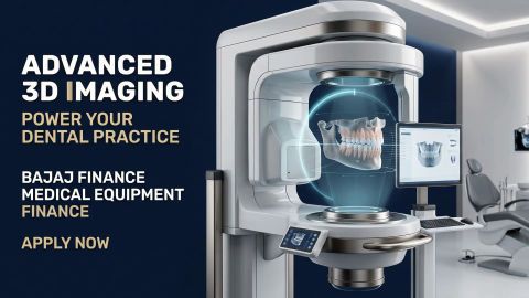
CBCT Scanner Uses Working and Price Guide
In summary
A CBCT scanner produces 3D images of teeth, jaws and other craniofacial structures. It helps clinicians assess selected dental and ENT conditions and plan appropriate treatments. Here are the key points to know:
- A CBCT scanner uses a cone-shaped X-ray beam and a rotating detector. Software combines the captured images into a 3D view.
- CBCT supports dental implant planning, orthodontics, endodontics and oral surgery. It also helps with selected ENT assessments.
- Small-FOV scanners capture focused dental areas. Larger-FOV scanners cover wider craniofacial regions.
- Prices depend on the brand, field of view, software and features. Installation and service support also affect the cost. New systems may cost around Rs. 15 lakh to Rs. 60 lakh or more.
- Compare image quality, radiation-dose optimisation and room requirements. Check the warranty, servicing and applicable AERB requirements.
- Eligible healthcare professionals and healthcare-facility promoters can explore Bajaj Finance Medical Equipment Finance.
Carefully assess your clinical requirements, installation costs and ongoing maintenance before choosing a CBCT scanner. Explore suitable financing options to plan your equipment investment.
Last updated: 13 October 2026
What is a CBCT scanner?
-
A CBCT scanner is a specialised imaging system that uses a cone-shaped X-ray beam to produce three-dimensional images of anatomical structures. CBCT is commonly used in dentistry to examine teeth, jaws, facial bones and surrounding structures in greater detail than conventional two-dimensional dental radiographs can provide. Depending on the scanner and its field of view (FOV), clinicians can assess a specific tooth, part of the jaw or a wider craniofacial region. These images can support diagnosis and treatment planning for selected dental and ENT conditions.
CBCT does not replace every type of medical imaging. The clinician should select the imaging method according to the patient's symptoms, clinical question and the expected diagnostic benefit.
How does a CBCT scanner work?
A CBCT scanner captures multiple two-dimensional X-ray images as the source and detector rotate around the patient. Specialised software then reconstructs these images into a three-dimensional representation.
The process generally involves the following steps:
- Patient positioning: The patient is positioned according to the scanner's instructions and the required scan area.
- Image capture: The X-ray source and detector rotate around the relevant anatomical region.
- Digital reconstruction: Software processes the captured projections to create a 3D image.
- Image review: The clinician examines the reconstructed images to assess the structures relevant to the diagnosis or treatment plan.
Scan time and image quality vary by model, scan settings and the area being examined. The operator should use an appropriate field of view and exposure settings to avoid unnecessary radiation.
What are the uses of a CBCT scanner?
CBCT imaging supports several dental and selected ENT applications where three-dimensional visualisation can help clinicians assess anatomical structures.
| Clinical application | How CBCT may help |
| Dental implant planning | Assesses jawbone dimensions and the position of relevant anatomical structures before implant placement |
| Endodontics | Helps evaluate complex root anatomy, suspected root fractures and selected periapical conditions |
| Orthodontics | Supports assessment of impacted teeth, jaw relationships and selected craniofacial structures |
| Oral and maxillofacial surgery | Helps with pre-operative assessment and planning for selected procedures |
| ENT assessment | Supports imaging of structures such as the paranasal sinuses and temporal bones, where clinically appropriate |
| Airway assessment | Provides anatomical information about the upper airway in selected clinical evaluations |
CBCT findings must be interpreted alongside the patient's symptoms, examination and other relevant clinical information. A scan alone does not confirm every suspected condition, including cancer.
What are the types of CBCT scanners?
CBCT scanners differ in their field of view, imaging capabilities and intended clinical applications. The field of view determines the anatomical area captured during a scan.
| CBCT scanner type | Typical field of view | Primary application |
| Dental implant CBCT | Small to medium | Jawbone assessment and implant planning |
| Endodontic CBCT | Small | Detailed imaging of individual teeth and root structures |
| Orthodontic CBCT | Medium to large | Assessment of jaw relationships, impacted teeth and craniofacial structures |
| ENT CBCT | Medium to large | Imaging of the paranasal sinuses, temporal bones and related structures |
The exact field-of-view dimensions and available features vary by model. Confirm the manufacturer's specifications before selecting a scanner.
How does the field of view affect CBCT scanner performance?
The field of view determines the size of the area captured in a scan. Selecting an appropriate field of view helps balance diagnostic requirements, image quality and radiation exposure.
| Field of view | Coverage | Common applications |
| Small FOV | A limited area, such as an individual tooth or local region | Endodontics and focused dental assessments |
| Medium FOV | A larger section of the jaw or dental arches | Implant planning and selected oral surgery assessments |
| Large FOV | A wider craniofacial region | Selected orthodontic, surgical and ENT assessments |
A larger field of view is not automatically better. Choose the smallest field of view that adequately answers the clinical question, following appropriate imaging protocols.
Get professional loan for your needs

Personal loan for doctors for wedding
Get doctor loan for for wedding

Chartered accountant loan
Get CA loan with minimal documents
CA loan for setting practice
Get CA loan for setting up your practice
CA loan for office renovation
Get CA loan for renovating office

Business loan for lawyer
Get business loan for lawyer today

Business loan for CA
Get business loan for CA today
Medical equipment finance for diagnostic centre
Setup diagnostic centre with medical equipment finance

Personal loan for lawyer
Get a personal loan for lawyer

Personal loan for doctors for wedding
Get a personal loan for wedding

Personal loan for doctors
Get a personal loan for doctor

Personal loan for CA
Get a personal loan for CA
Medical equipment finance
Get a medical equipment finance

Lawyer loan to start firm
Get a lawyer loan to start firm

Lawyer loan for Chamber
Get a lawyer loan for Chamber

Lawyer loan
Get a lawyer loan

Hospital loan
Get a hospital loan

Doctor loan for setting up practice
Get a doctor loan for setting up practice
Doctor loan for clinic renovation
Get a doctor loan for clinic renovation

Doctor loan
Get a doctor loan
What is the difference between a CBCT scanner and an OPG machine?
An orthopantomogram (OPG) machine produces a two-dimensional panoramic dental image, while a CBCT scanner reconstructs three-dimensional images from multiple X-ray projections.
| Feature | OPG machine | CBCT scanner |
| Image type | Two-dimensional panoramic image | Three-dimensional reconstructed image |
| Anatomical detail | Provides a broad dental overview | Provides detailed views of selected anatomical structures |
| Common applications | Routine dental assessment and selected orthodontic evaluations | Implant planning, selected endodontic assessments and complex surgical planning |
| Measurements | Limited depth information | Enables three-dimensional measurements |
| Radiation exposure | Generally lower than CBCT | Varies by field of view, equipment and protocol; generally higher than conventional dental radiography |
| Equipment investment | Usually lower | Usually higher |
Neither system is suitable for every diagnostic requirement. The clinician should select the imaging method that provides sufficient information for the clinical question while avoiding unnecessary exposure.
How can you finance a CBCT scanner with Medical Equipment Finance?
Purchasing a CBCT scanner can require a substantial upfront investment. Eligible doctors and healthcare facilities can explore Bajaj Finance Medical Equipment Finance to finance eligible medical equipment purchases.
Key product details include:
- Loan amount: Rs. 2 lakh to Rs. 30 crore.
- Repayment tenure: 3 to 120 months.
- Funding: Financing of up to 100% of the equipment value
- Approval: Within 48 hours* of disbursal
Before applying, estimate the equipment cost, installation expenses and expected operating cash flow. Compare the repayment schedule with your income to assess whether the proposed borrowing fits your budget.
Planning to upgrade your healthcare facility or purchase medical equipment? Check your eligibility for Medical Equipment Finance from Bajaj Finance and explore funding options for eligible equipment purchases, subject to eligibility and applicable terms.
Our loan variants
Use our popular calculators






Types of medical equipment loan

Business loan for doctors

Business loan for chartered accountants

Set up a diagnostic centre with medical equipment finance

How to start a law firm in India

Doctor loan for clinic renovation
Frequently asked questions
Understanding CBCT scanners
Price and purchasing considerations
Clinical use and financing
What is the full form of CBCT?
CBCT stands for Cone Beam Computed Tomography. It is an imaging technology that uses a cone-shaped X-ray beam and a rotating detector to produce three-dimensional images. In dentistry, it helps clinicians assess teeth, jaws and surrounding anatomical structures for selected diagnostic and treatment-planning requirements.
Can CBCT scanner detect cracked tooth?
A CBCT scan may help identify certain dental fractures that are difficult to assess on conventional radiographs. However, the ability to detect a crack depends on its size, orientation, location, image quality and other clinical factors. A dentist must interpret the scan alongside the examination and other findings. A CBCT scan cannot reliably identify every crack.
What is the average lifespan of a CBCT scanner?
The average lifespan of a CBCT scanner is around 10–15 years, depending on usage, maintenance, software updates, and manufacturer quality, ensuring long-term value for medical and dental practices.
Is a refurbished CBCT scanner cheaper than a new scanner?
A refurbished CBCT scanner may have a lower purchase price than a comparable new model. However, the total cost depends on its age, condition, software, warranty, servicing requirements and regulatory status.
Why do large-FOV CBCT scanners cost more than small-FOV models?
Large-field-of-view scanners capture a wider anatomical area and may include more advanced imaging capabilities or integrated functions. These features can increase the equipment cost. However, the most appropriate scanner depends on your clinical requirements, not simply the size of its field of view.
How does the field of view affect CBCT scanner performance?
The field of view determines the anatomical area captured in a scan. Small-FOV systems are useful for focused dental imaging, while medium- and large-FOV systems can cover wider regions for selected implant, orthodontic, surgical and ENT applications. The appropriate FOV depends on the clinical question, required image detail and radiation protection considerations.
How does field of view (FOV) affect a CBCT machine's performance?
The field of view (FOV) determines the size of the anatomical area captured in a single scan. Small FOV provides higher image resolution for endodontics and implant planning, while medium and large FOV support orthodontics, oral surgery, and ENT imaging by covering larger regions with fewer scans and greater diagnostic versatility.
What is the ROI on purchasing a CBCT machine for a dental clinic in India?
The ROI on a CBCT machine depends on scan volume, pricing, and financing costs. Clinics performing implantology, orthodontics, and oral surgery can often recover their investment within 18–30 months through consistent patient demand. In-house CBCT imaging also increases treatment acceptance, reduces referrals, and generates an additional recurring revenue stream.
What do our customers say about us
Dr. Rohan Mehta
Reviewed on
I applied for a doctor loan to upgrade my clinic infrastructure, and the experience was excellent. The application process was simple, the documentation was minimal, and the approval came through quickly. The team guided me throughout the process and ensured a seamless experience. I would highly recommend this financing option to medical professionals looking for reliable funding support.
Dr. Priya Sharma
Reviewed on
The doctor loan helped me manage my professional expenses without any financial stress. The loan disbursal was fast, and the customer support team was always available to answer my questions. The repayment options were flexible and suited my financial requirements perfectly. Overall, I am very satisfied with the service and support provided.
Dr. Arvind Nair
Reviewed on
I availed a doctor loan to purchase advanced medical equipment for my practice. The entire process was smooth, transparent, and completed within a short time. The competitive interest rates and hassle-free documentation made the experience even better. The support team remained helpful at every stage, making the loan journey convenient and stress-free.
Dr. Neha Kapoor
Reviewed on
I needed financial assistance to expand my healthcare practice, and this doctor loan proved to be the right solution. The approval process was quick, and the team provided clear guidance from application to disbursal. The flexible repayment tenure helped me plan my finances effectively. I am pleased with the overall experience and would recommend it to fellow doctors.
More articles

Why choose Bajaj Finance medical equipment finance?
Read More
6 benefits of medical equipment loans
Read More
Set up a diagnostic centre with Medical Equipment Finance
Read More

Role of credit score in securing medical equipment financing
Read More
Disclaimer
Bajaj Finance Limited has the sole and absolute discretion, without assigning any reason to accept or reject any application. Terms and conditions apply*.